Fig. 1
From: Lumbar vertebral canal stenosis due to marked bone overgrowth after routine hemilaminectomy in a dog
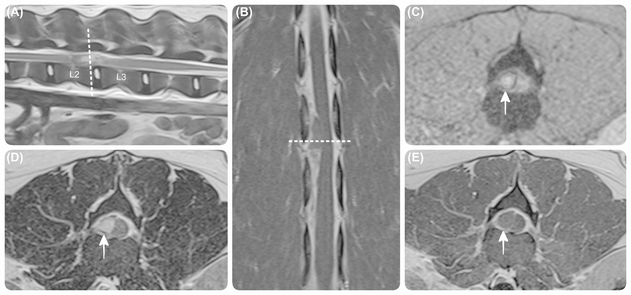
Comparison between different magnetic resonance images at the level of L2-L3 vertebrae. Sagittal T2W (A), dorsal T1W post-contrast (B), and transverse T2*GRE (C), T2W (D), T1W post-contrast (E) magnetic resonance images at the level of L2-L3 vertebrae, showing a right-sided extradural lesion (white arrow), extending from mid-L2 to mid-L3 vertebral body levels. The lesion showed heterogeneous, hyperintensity on T2W and T1W images, and had a complete hypointense rim and hyperintense centre on T2*-GRE images. The lesion exhibited faint peripheral contrast enhancement.