Fig. 2
From: Lumbar vertebral canal stenosis due to marked bone overgrowth after routine hemilaminectomy in a dog
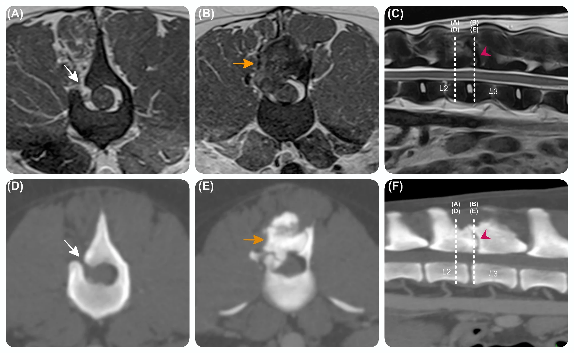
Advanced diagnostic imaging performed a year post-routine hemilaminectomy. Transverse T2W (A) (B), and sagittal T2W (C) magnetic resonance images and transverse (D) (E) and sagittal (F) computed tomography images at the level of the L2-L3 vertebrae, showing a relatively sharp and small laminar defect (H 1 mm x L 4.5 mm) (white arrow) on the right side of caudal aspect of L2 vertebra. Adjacent to this site and extending caudally, there was an ill-defined expansive lesion (yellow arrow) lesion arising from the vertebra, affecting the right articular processes, and ventral aspect of the spinous process of L2-3 and the contiguous vertebral laminae, causing mild spinal cord compression. The lesion was slightly heterogeneous in all sequences, with mild heterogeneous contrast enhancement.