Fig. 3
From: Lumbar vertebral canal stenosis due to marked bone overgrowth after routine hemilaminectomy in a dog
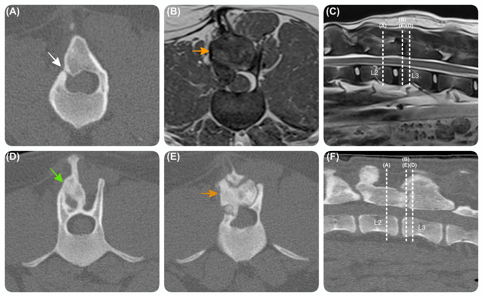
Advanced diagnostic imaging performed 9 months post-revision surgery. Transverse (A) (D) (E) and sagittal (F) computed tomography images and transverse T2W (B) and sagittal (C) magnetic resonance images at the level of the L2-L3 vertebrae, showing a right-sided expansive lesion (orange arrow) similar to the Fig. 2, with smooth bone almost completely filling the previous laminectomy defect, but leaving a small laminar gap (H 1 mm x L 1 mm), on the right side of the caudal aspect of L2 vertebra (white arrow). The lesion has grown more eccentrically, and incorporates the slightly deviated L2 and L3 (green arrow) spinous processes.